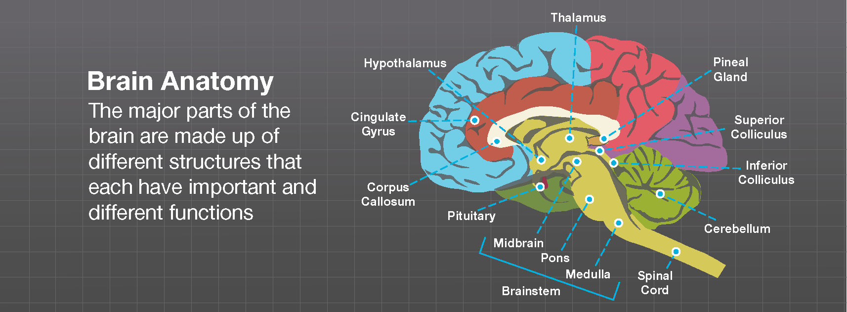

However, the structures within the MB-HB are small and move with cardiac pulsations therefore, physicians were slow to recognize subtle distortions in their structure and recognize their importance in developmental disorders. The ability to acquire high resolution, high contrast, distortion-free images in sagittal and coronal planes allowed accurate gross assessment of MB-HB structures for the first time. Malformations of the MB or HB secondary to defects in anteroposterior (AP) or dorsoventral (DV) patterning were nearly impossible to identify in neurology patients until magnetic resonance imaging became a commonly used tool in clinical diagnosis.

Malformations secondary to early patterning defects This review will highlight a few MB-HB malformations and emphasize how knowledge of basic research in embryology, genetics, and cellular and molecular biology of the developing brain can be of importance in recognizing, understanding, and classifying these anomalies in humans. These disorders were recently extensively reviewed and classified (Barkovich et al., 2009). The number and complexity of recognized malformations of the brainstem and cerebellum has been steadily increasing. Among the most common of these are lissencephalies (Ross et al., 2001 Lecourtois et al., 2010), so-called “cobblestone malformations” of the cortex (formerly known as lissencephaly type II) resulting from defects in the pial limiting membrane (van Reeuwijk et al., 2006 Clement et al., 2008 Hewitt, 2009), anomalies of the cerebral commissures (Barkovich et al., 2007), and disorders of primary cilia function that include additional ocular, renal, hepatic, and limb bud anomalies (Lancaster et al., 2011 Sang et al., 2011). Although malformations of the brainstem and cerebellum may be the only recognized abnormality in individuals with mental retardation or autism (Soto-Ares et al., 2003 Courchesne et al., 2005), they are more commonly identified in patients with malformations of the cerebrum. Recently, however, advances in developmental genetics, neurobiology, molecular biology, and neuroimaging have led to better understanding of developmental disorders of the embryonic midbrain (MB) and hindbrain (HB), which grow into the adult brainstem and cerebellum (Barkovich et al., 2007, 2009). Radiologic analysis of these structures by pneumography, angiography, and X-ray computed tomography was poor. The cerebellum was believed to have a minor role in brain function, while the brain stem was difficult to remove intact at autopsy and difficult to section. For many years, anomalies of the cerebellum and brain stem were poorly reported in the scientific literature.


 0 kommentar(er)
